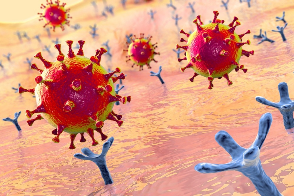
In a recent study posted to the bioRxiv* preprint server, researchers evaluated the use of receptor decoys for severe acute respiratory syndrome coronavirus 2 (SARS-CoV-2) based on soluble forms of angiotensin-converting enzyme 2 (ACE2) for immunological prophylaxis and management of coronavirus disease 2019 (COVID-19) through the use of adeno-associated virus (AAV) vectors.

Background
Vectored immunological prophylaxis for SARS-CoV-2 through the expression of neutralizing monoclonal antibodies or vaccines is challenging due to the rapid evolution of SARS-CoV-2. ACE2 receptor decoys may inhibit SARS-CoV-2 entry and are less susceptible to immune escape by novel SARS-CoV-2 variants of concern (VOCs).
The authors of the present study previously developed an ‘ACE2 microbody’ receptor decoy protein by fusing the ACE2 ectodomain to the CH3 domain of a human immunoglobulin 1 (IgG1) heavy chain fragment crystallizable (Fc) region. Intranasal (i.n.) administration of the microbody protected against infection in hamsters and mice when administered shortly before infection and suppressed SARS-CoV-2 replication when administered after 12.0 hours of infection.
About the study
In the present study, researchers applied the vectored immunological prophylaxis concept to establish long-term COVID-19 prophylaxis by lentiviral and AAV vectors expressing a modified high-affinity ACE2 microbody.
The feasibility of vectored immunological prophylaxis against SARS-CoV-2 was determined by constructing AAV vectors expressing the ACE2.1mb decoy receptor of ACE2. The decoy protein was altered by inserting point mutations in the site of ACE2-SARS-CoV-2 S (spike) protein binding to enhance S protein binding affinity and by inserting the H345A point mutation for catalytic activity inactivation. Coding sequences were cloned into AAV vectors comprising CAG promoters, and SARS-CoV-2 stock was produced with AAV6.2 and AAV2.retro capsids.
The protective ability of the vectors against SARS-CoV-2 infection was evaluated using the A549.ACE2 lung cells and the CHME3.ACE2 microglial cells. The cells were transduced with the decoy vectors and challenged five days later with the SARS-CoV-2 D614G strain and Omicron sub-VOCs, including the BA.1 sub-VOC, BA.2 sub-VOC, BA.2.75 sub-VOC, BA.4 sub-VOC, BA.5 sub-VOC and the BQ.1 sub-VOC S protein-pseudotyped lentiviruses.
Luciferase activity in cell cultures was evaluated at 2.0 days post-infection (dpi). The ability of the decoy-expressing vectors to inhibit SARS-CoV-2 live virus replication was tested on A549.ACE2, CHME3.ACE2 and the hSABCi-NS1.1 human small airway basal cells.
SARS-CoV-2 proliferation was measured by quantifying SARS-CoV-2 RNA copies by quantitative reverse-transcription-polymerase chain reaction (RT-qPCR) analysis. The transduced cells were analyzed by flow cytometry to discriminate between the types of pulmonary cells. The practicality of the vectored COVID-19 prophylaxis was determined in vivo by administering ACE2.1mb-expressing AAV vectors to ACE2 K18 transgenic mice intramuscularly or intranasally, followed by SARS-CoV-2 WA1/2020 challenge, and pulmonary viral load assessments at 3.0 dpi.
Antibodies eBook

The efficacy of decoy vectors in protecting against the Omicron sub-VOCs was assessed using the BALB/c murine model. The durability of SARS-CoV-2 suppression was determined by challenging vector-treated mice with SARS-CoV-2 over 30 days. The protective efficacy of the vectors was compared to that of the highly potent LY-CoV1404 monoclonal antibody.
Results
Intramuscular or intranasal administration of ACE2.1mb-expressing AAV6.2 and AAV2.retro vectors conferred a high degree of protection against COVID-19 in BALB/c mice and ACE2 K18 transgenic mice. Lentiviral- and AAV-vectored immunological prophylaxis were durable and effective against the recently emerged Omicron sub-VOCs. In addition, the vectors showed therapeutic efficacy when delivered ≤1.0 dpi, with non-diminishing suppression of SARS-CoV-2 replication by the lentiviral vector-based decoy for ≤2.0 months post-intranasal instillation. LY-CoV1404 was less effective than the lentiviral vectors in reducing viral loads.
D614G and Omicron BA.5 were most potently neutralized by the decoy vectors, whereas Omicron BA.2 was the most resistant, with 20-fold to 33-fold greater ID50 values, similar to the in vitro findings. The transduced CHME3.ACE2 cells with the AAV vectors resulted in a four-log to a five-log reduction in SARS-CoV-2 RNA, a level not significantly greater than that observed in the uninfected cells. Similar findings were obtained using A549.ACE2 cells. The AAV vectors were also effective in the hSABCi-NS1.1 cells, albeit the reduction was less prominent (50-fold to 100-fold).
Histopathological examination of pulmonary tissues of ACE2.1mb-expressing vectors-treated mice showed no interstitial pneumonia-like features and lack of inflammatory cell infiltration. In addition, cytokines such as interferon-alpha (IFN-α), interleukin 6 (IL-6), tumor necrosis factor-alpha (TNF-α), IL-10, monocyte chemoattractant protein 1 (MCP-1), and IL12-p70 were not induced following vector or decoy administration. Moreover, the decoy vectors prevented weight loss in the animals.
Intramuscular delivery of AAV vectors was more effective at reducing the viral loads than intravenous administration (five-log reduction vs. two-log reduction). The findings indicated that long-term lentiviral vector expression resulted from elevated leukocyte and dendritic cell transduction. Intravenous administration of the lentiviral vector resulted in elevated expression in the spleen and moderate levels in the liver and lungs. Intranasal administration resulted in elevated levels in the lungs and moderate expression in the nasal tissues and trachea.
Conclusion
Overall, the study findings showed that vectored immunological prophylaxis could be highly useful to confer rapid immune protection among immunosuppressed individuals for whom vaccination is not practical or less effective against recently emerged SARS-CoV-2 variants.
*Important notice
bioRxiv publishes preliminary scientific reports that are not peer-reviewed and, therefore, should not be regarded as conclusive, guide clinical practice/health-related behavior, or treated as established information.
- Tada, T. et al. (2023) "Vectored Immunoprophylaxis and Treatment of SARS-CoV-2 Infection". bioRxiv. doi: 10.1101/2023.01.11.523649. https://www.biorxiv.org/content/10.1101/2023.01.11.523649v1
Posted in: Medical Science News | Medical Research News | Disease/Infection News
Tags: ACE2, Angiotensin, Angiotensin-Converting Enzyme 2, Antibodies, Antibody, binding affinity, Cell, Coronavirus, Coronavirus Disease COVID-19, covid-19, Cytokines, Cytometry, Dendritic Cell, Efficacy, Enzyme, Evolution, Flow Cytometry, Immunoglobulin, in vitro, in vivo, Interferon, Interleukin, Leukocyte, Liver, Luciferase, Lungs, Monoclonal Antibody, Monocyte, Mutation, Necrosis, Omicron, Pneumonia, Point mutation, Polymerase, Polymerase Chain Reaction, Proliferation, Prophylaxis, Protein, Receptor, Respiratory, RNA, SARS, SARS-CoV-2, Severe Acute Respiratory, Severe Acute Respiratory Syndrome, Spleen, Syndrome, Transcription, Transgenic, Tumor, Tumor Necrosis Factor, Virus, Weight Loss

Written by
Pooja Toshniwal Paharia
Dr. based clinical-radiological diagnosis and management of oral lesions and conditions and associated maxillofacial disorders.
Source: Read Full Article
