
There’s been an explosion in the use of endoscopes in medical procedures over the past 30 years, from examining the liver and stomach (known as laparoscopy) to minimally invasive surgery. The use of these instruments is firmly established as an imaging technique that allows surgeons to ‘see’ inside the body, allowing them to recognize lymph nodes they need to avoid, or assess the health of lung tissue, for example.
One of the most advanced types of endoscopes relies on bundles of minuscule optical fibers—usually thousands of them—to give doctors a picture of the area of concern, and they have saved countless lives.
While optical fibers are perfect for the role—being thin, flexible, chemically inert and non-toxic, as well as immune to electromagnetic interference from other medical instruments—they have one disadvantage: doctors can only see in two dimensions, and this flatness of image is a limitation when diagnosing conditions. But that’s all about to change.
Researchers at the Centre for Nanoscale BioPhotonics (CNBP) have developed a clever technique to make the tiny, sometimes blurry, images of cellular regions not only sharper but 3-dimensional. The research has the potential to improve endoscopy by providing a higher quality and more detailed image.
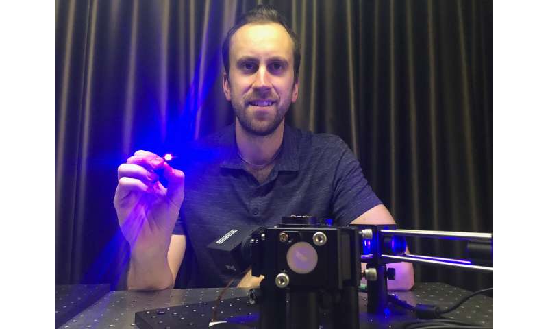
“We went back over the physics of how the optical fiber bundles work and realized that they actually transmit 3-D images as well,” said Dr. Antony Orth, a former research fellow at CNBP’s RMIT University node in Melbourne under Prof Brant Gibson, CNBP chief investigator for nanomaterials and multimodal imaging.
“People generally assume that each fiber in the bundle only reports the brightness at the other end. But there’s more to it. If you send light at different angles into the fiber, it exits in these beautiful patterns called modes,” he added. “That’s where the magic happens.”
Dr. Orth had been reviewing the basic physics to look for new uses and exchanging ideas around imaging with CNBP colleague Dr. Martin Plöschner of the University of Queensland.
“After playing around with these fibers for a few months, it became apparent that if you shine a light at one end at a really high angle, at the other end you see all these crazy patterns, just seemingly randomly oriented,” said Dr. Orth. “It’s a really striking visual effect and totally different to what you get when using the fiber under normal circumstances.
“I realized that we weren’t just seeing spatial distribution of light, we were also seeing the angular distribution—and if you can measure both of those at the same time, you have a ‘light field,”‘ he added.
As it turned out, Dr. Orth’s doctorate had centered on light fields, and he could immediately see how the spatial and angular information could be combined to create a 3-D image. “You can reorganise the light rays to change focus, or send one group of angles to one sensor and another group to another, and have a stereoscopic image,” he said. “It’s like having a tiny multifocal lens in the fiber.”
But making it a reality was anything but straightforward. “I knew we should be able to do this, but it took another couple of years to be able to fully realize it,” he said. “We had to flesh out the math to be able to quantitatively turn the modal information into depth information, which was the most challenging part. The intuition was there, you just have to keep slaving away at the math.”
The size of microendoscopes makes them perfect for accessing hard-to-reach areas of the human body. But until now, they’ve been too small to contain the elements needed for complex focusing optics. But ‘light field imaging,” as the technique is known, allows focusing, stereo visualization and depth mapping at the fiber’s tip, relying on optical fiber bundles already in clinical use. The bundles are just three-quarters of a millimeter wide and contain 30,000 fibers.
“It gives you the 3-D structure of whatever tissue you’re looking at, which may allow you to tell whether a tissue is benign or malignant or somewhere in between,” said Dr. Orth. “We’ve experimentally demonstrated you can get 3-D information this way. The next step is doing it in animals and then humans.”
In fact, the team’s approach to microendoscopic light field fluorescence imaging—for which a patent is pending—may establish optical fiber bundles as a new class of light field sensor, able to see surface features and map their depth with a rate of accuracy previously unobtainable. And there’s an added advantage that the technique uses off-the-shelf fibers, which will hopefully translate to faster adoption in medicine. “The idea is to try to get the same sort of information you get from a biopsy, but do it right at the site. That’s what we want to work towards. This is the first step toward that.”
Source: Read Full Article