The human knee joint is the largest and most complex joint in the body. It has combines hard and soft tissue elements to provide stability and proper functioning, including the meniscus. The meniscus, a cartilage pad between the thighbone and the shinbone, acts as a shock absorber to dissipate the body weight and reduce friction during physical activities. Considering the knee joint is the most-used joint in the body, meniscal tears are the most common knee injury due to wear and tear due to age and injuries.
Meniscal tears can be treated via physiotherapy, surgery to remove the torn tissue, replacement with allografts, or with implants. While meniscal transplants serve as an alternative option, it is technically challenging due to the customization in the sizing required for each person, including positioning, securing the transplant to the bone, and the risks of rejection and infection.
Even though a significant amount of work and research has been carried out on prostheses and implants, meniscal implants in particular seem to have been significantly overlooked. It was only in 2015 that the first meniscal replacement was implanted in humans during a clinical trial, and in September 2019, the U.S. Food and Drug Administration approved the first artificial meniscus based on clinical trial results.
To bridge this research gap, Singapore University of Technology and Design’s (SUTD) Medical Engineering and Design (MED) Laboratory collaborated with the University of Miyazaki’s Biomechanics Laboratory and developed a novel methodology for the early stage design assessment and verification of meniscal implants using computational modeling and simulation. This initial design-stage development paves the way for the manufacture of a customized 3-D printed meniscus for a more optimal fit. The research paper has been published in IEEE Access.
This new development will provide medical practitioners and industry experts with valuable insights into how the remnant hard and soft tissues in the knee joint react biomechanically to an artificial meniscus. They can then use this information to evaluate aspects of in vivo performance without subjecting patients or animals to potential harm or unnecessary risk. Also, the computational models can be used to evaluate options that would otherwise have been impossible experimentally or clinically.
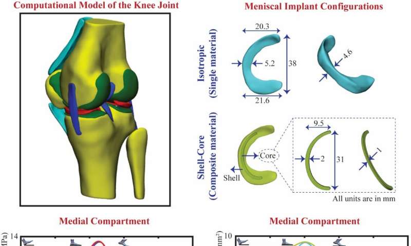
To demonstrate this, the team developed a comprehensive and detailed computational model of an intact knee joint from the medical images and gait cycle data of a healthy adult volunteer. They validated the developed model with biomechanical experiments using the lower extremity of cadavers. They used the validated model to analyze the biomechanical response of soft tissues in the knee joint for different meniscus conditions, such as intact meniscus, meniscal root tear, total removal of the meniscus, single-material meniscal implant and a shell-core composite meniscal implant. Their results suggested that the composite meniscal implant with a shell-core structure performed better than other meniscus conditions and restored the natural biomechanical response of the joint.
This study could also explain the development of knee osteoarthritis due to increased contact stresses and altered joint kinematics caused by the loss of meniscal tissue. This novel synthetic meniscal implantation approach restores the joint mechanics close to the intact meniscus state and appears to be a promising strategy for treating patients with severe meniscal injuries. The model developed in this study sheds light on the knowledge of joint mechanics after injury or repair, and therefore can also assist in the clinical evaluation of other alternative repair techniques.
Source: Read Full Article
