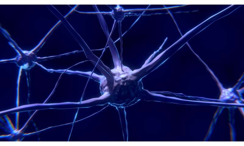
Like buoys bobbing on the ocean, many receptors float on the surface of a cell’s membrane with a part sticking above the water and another underwater, inside the cell’s cytoplasm. But for cells to function, these receptors must be docked at specific regions of the cell. Most research has focused on the ‘underwater’ portions. That’s where the cell’s molecular machines swarm and interact with a receptor’s underwater tails, with those interactions then fueling signals that dive deep into the nucleus, changing the cell’s course.
New work by a team of Thomas Jefferson University researchers reveals new activity above the surface, in brain-cell receptors that govern learning and chronic pain. In the study, the authors show that the ‘above water’ portion of proteins can help dock the proteins at synapses, where neurons mediate flow of information throughout the brain. This discovery opens the possibility of using this docking site as a target to develop treatments for chronic pain and other diseases more effectively. The study was published January 29th in Nature Communications.
“The extracellular spaces—the parts ‘above the water’ – have been largely overlooked,” says senior author Matthew Dalva, Ph.D., professor and vice chair of the Department of Neuroscience and director of the Jefferson Synaptic Biology Center in the Vickie & Jack Institute for Neuroscience – Jefferson Health. Dr. Dalva and his team looked at the NMDAR receptor on brain cells and pinpointed the spot where this receptor interacts with a neighbor to initiate signaling. “When trying to develop new therapy, finding the bullseye is half the problem,” says Dr. Dalva.
Finding a key interaction that sits above the cell’s surface, could make it more accessible to therapeutics. “The kinds of receptor interactions we’re talking about are different than when a receptor binds to its ligand outside of the cell, which is well documented,” says Dr. Dalva. “Here we’re describing the kinds of biochemical exchanges—kinase phosphorylation fueled by free-floating ATP—that we thought, until recently, were exclusive to the inside of cells.”
The researchers focused on the synaptic protein called NMDA-type glutamate receptors (NMDARs), which help regulate the strength of synaptic connections between neurons. It’s important that the synapse connects strongly, but not too strongly, in order to prevent creating an overly excitable connection.
A key mechanism controlling synaptic strength is the increase in NMDAR function due to direct molecular interaction with another synaptic protein called the EphB receptor tyrosine kinase. Dr. Dalva and colleagues had previously shown that the phosphorylation of EphB on the “outside” or extracellular portion of the molecule can lead to a direct interaction with NMDARs. And that chemical exchange causes the receptors to cluster and drive neuronal plasticity and chronic pain (PlosBiology 2017). Their new work identifies a specific region of the NMDAR or bullseye, necessary for these proteins to interact.
This specific bullseye could have important medical implications, as disruption of the EphB-NMDAR interaction has been associated with Alzheimer’s, and in chronic pain can be due to too much of this interaction. As a trans-synaptic organizer and NMDAR binding protein, the EphB receptor is a key regulator of these events.
However, despite discovery of this interaction over a decade ago, the exact spot where NMDAR interacts with EphB has been a mystery. Here, the researchers demonstrate that specific amino acids in the hinge region of the NDMAR are required to interact with EphB2. Importantly, the amino acids in the hinge region are required for proper NDMAR mobility and stabilization at the synapse.
Source: Read Full Article
