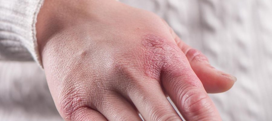
Throughout the coronavirus disease 2019 (COVID-19) pandemic, there have been increased reports of chilblain lesions in individuals with an active severe acute respiratory syndrome coronavirus 2 (SARS-CoV-2) infection. Affected adolescents and young adults with no comorbidities would present with swollen, erythematous, tender macules and papules, as well as ulcers on hands and/or feet, thus leading many healthcare providers to suspect SARS-CoV-2 as the source of these lesions.
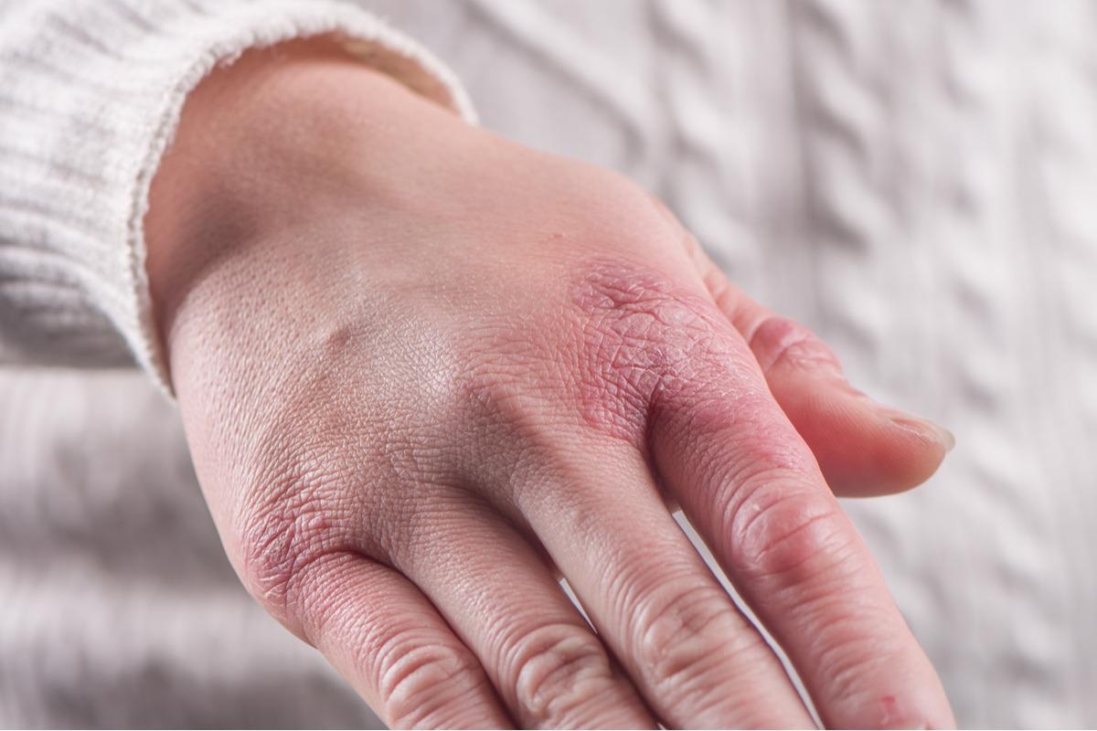
Study: Pandemic chilblains: Are they SARS-CoV-2-related or not? Image Credit: kungfu01 / Shutterstock.com
Background
Chilblains are lesions often induced by cold weather that can lead to anoxia, capillary injury, and dermal inflammation as a result of vasoconstriction of the deep cutaneous arterioles. Both coagulopathy and autoimmune disorders have been linked to the formation of chilblain lesions.
Chilblain lesions that have been detected throughout the pandemic do not differ clinically or histopathologically from those seen before the pandemic, with the exception of a possibly more severe feature in the pandemic group.
In a recent Clinical Immunology review, researchers evaluate the pathophysiological relationship between COVID-19 and pandemic-associated chilblain lesions. Furthermore, the authors of this review determine whether cold exposure, coupled with a lockdown-imposed sedentary lifestyle, is an alternative explanation for chilblain lesions during the current pandemic.
Chilblains and COVID-19 peaks
SARS-CoV-2 has been considered the primary source of chilblains during the current pandemic, as it has been shown to induce lesions in a variety of tissues. Thus, researchers have questioned whether chilblains are more common in places with a high COVID-19 prevalence.
Chilblains were most often observed during COVID-19 lockdowns. Several studies found associations between chilblains and lockdown circumstances in their respective nations, individual lifestyle changes, garment conditions, cold exposure, and recognized chilblain risk factors including a higher body mass index (BMI).
Despite infection rates that were similar to those reported in nations with a higher incidence of chilblains, no chilblain outbreaks occurred in Nordic countries nor tropical countries. Chilblain outbreaks were reported in Belgium during the first and second COVID-19 waves, which were marked by lockdowns.
However, proceeding COVID-19 waves that occurred after lockdown restrictions were lifted were associated with chilblain lesions rates that were similar to those observed in previous years. These epidemiological findings demonstrate that the greater incidence of lesions reported in patients with known or unknown risk factors for chilblains could be associated with lockdown-imposed sedentary lifestyles and extended barefoot contact on cold floors.
SARS-CoV-2 and chilblain correlation at the individual level
Patients with chilblain lesions showed no or mild symptoms consistent with SARS-CoV-2 infection. Anti-SARS-CoV-2 antibodies and reverse transcription-quantitative polymerase chain reaction (RT-qPCR) on nasopharyngeal swabs were negative in the majority of patient series, whereas anti-SARS-CoV-2 antibodies were positive in the majority of control patients with PCR-confirmed infection.
Several groups used antibodies to identify SARS-CoV-2 in chilblain tissue sections. The findings are incongruent, thereby indicating that existing immunostainings for SARS-CoV-2 in the skin are insufficiently reliable.
SARS-CoV-2 was also not detected using RT-PCR on isolated ribonucleic acid (RNA) material or RNAscope in situ hybridization from chilblain tissue samples. Overall, the evidence does not support the hypothesis that SARS-CoV-2 is the direct cause of all pandemic chilblains.
Spatial and temporal association of SARS-CoV-2 and chilblains
Despite the viral and antibody undetectability in symptomatic patients, the spatial and temporal correlation of SARS-CoV-2 with chilblains led to a theory involving type I interferons (IFN-I). IFN-I is produced by infected cells and can inhibit virus development or cause apoptosis in infected adjacent cells. Notably, COVID-19 patients with reduced IFN-I expression, either constitutively or as a result of anti-IFN autoantibodies, displayed greater disease severity.
The proposed theory for pandemic chilblains is that certain people have an unusually powerful IFN-I response, which destroys the virus before symptoms and apparent antibody responses occur, thus causing chilblains. It should be noted that this hypothesis has not been supported by substantial evidence. In fact, only a few researchers have found that IFN-I levels are elevated systemically, and only in a small number of patients with pandemic chilblains.
However, there is no evidence that people with pandemic chilblains have higher levels of IFN-I in their blood than those with seasonal chilblains. In fact, the transcriptome profiles of pandemic and seasonal chilblains were found to be identical.
If constitutively increased levels of IFN-I were responsible for pandemic chilblains, some of the systemic symptoms seen in interferonopathies would be expected. Furthermore, if a strong IFN-I response kills SARS-CoV-2 promptly at the start of the illness, viral proteins should not be detected weeks later.
Implications
The cause of pandemic chilblains has yet to be identified; however, SARS-CoV-2 should not be completely ruled out as a probable cause of this delayed symptom. Furthermore, further research must also be conducted to determine in SARS-CoV-2 can also trigger classic cold-related chilblains, which had a higher occurrence during the pandemic period. This may have been due to more prolonged and insidious cold exposure in a large number of patients confined to extraordinary confinement measures.
SARS-CoV-2 or other viruses, as well as genetic polymorphisms, could all contribute to the occurrence of chilblains lesions. Thus, a better understanding of the patients' environment in the weeks leading up to chilblains is required. More advanced technologies, like spatial transcriptomics or proteomics, can help to corroborate theories or uncover new mechanisms.
- De Greef, A., Coulie, P. G., & Baeck, M. (2022). Pandemic chilblains: Are they SARS-CoV-2-related or not? Clinical Immunology. doi:10.1016/j.clim.2022.108984.
Posted in: Medical Research News | Medical Condition News | Disease/Infection News
Tags: Adolescents, Anoxia, Antibodies, Antibody, Apoptosis, Autoantibodies, Blood, Body Mass Index, Chilblains, Cold, Coronavirus, Coronavirus Disease COVID-19, covid-19, Genetic, Healthcare, Hybridization, Immunology, Inflammation, Interferons, Nasopharyngeal, Pandemic, Polymerase, Polymerase Chain Reaction, Proteomics, Research, Respiratory, Ribonucleic Acid, RNA, SARS, SARS-CoV-2, Severe Acute Respiratory, Severe Acute Respiratory Syndrome, Skin, Syndrome, Transcription, Transcriptomics, Virus
.jpg)
Written by
Colin Lightfoot
Colin graduated from the University of Chester with a B.Sc. in Biomedical Science in 2020. Since completing his undergraduate degree, he worked for NHS England as an Associate Practitioner, responsible for testing inpatients for COVID-19 on admission.
Source: Read Full Article