
Both fluorescence microscopy and light microscopy represent specific imaging techniques to visualize cells or cellular components, albeit with somewhat different capabilities and uses. At its core, fluorescence microscopy is a form of light microscopy that uses many extra features to improve its capabilities.
 Vshivkova | Shutterstock
What is light microscopy?
Light microscopy does much what the name implies: visible light and magnifying lenses are used to view small objects. Light microscopes are the oldest form of higher quality imaging devices, dating back to the 1500s, and were the microscopes with which first cells were observed.
A common method to visualize cells or tissue with light microscopes is to use dyes. Widely used ones might paint the main components, such as the dye combination of hematoxylin and eosin, which colors the nuclei violet and the cytoplasm pink. However, there are also more specialized dye techniques.
In hospitals, quick examination of cells can be critical for doctors. In such situations, light microscopy can be used with tissues that have been frozen in carbon dioxide and sectioned using a microtome. This simpler method can be used urgently on patients who are in the operating room to guide the surgeon.
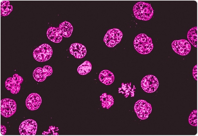
What is fluorescence microscopy?
As light microscopy developed, more forms using different techniques were invented. One of the types of microscopy within the broader light microscopy group is fluorescence microscopy. Fluorescence microscopy images cells or molecules that have been tagged with a fluorescent dye. The fluorescent substances are called fluorophores, which are integrated into the sample.
The fluorophores are excited by the light in the microscope, which causes them to give off light with lower energy and of longer wavelength. It is this light that produces the magnified view, rather than the original light source. This means that fluorescent microscopy uses reflected rather than transmitted light.
For example, a commonly used label is green fluorescent protein (GFP), which is excited with blue light and emits green light with a longer wavelength. Filters around the sample can separate the fluorescent light from the surrounding radiation.
Fluorescence microscopy is often applied in imaging cell structures or structural features, checking the viability of cells, imaging genetic material (both DNA and RNA), and imaging particular cells in a larger population.
Comparing light microscopy and fluorescence microscopy
As mentioned, light microscopes that are used for light microscopy employ visible light to view the samples. This light is in the 400-700 nm range, whereas fluorescence microscopy uses light with much higher intensity.
The usefulness of traditional light microscopy is hampered by the fact that it uses visible light, as using visible light limits the resolution obtained from samples. On the other hand, fluorescence microscopy is not faced with this limitation, since it uses whatever light excites the fluorophores.
Traditional light microscopes are widely used, and often require simpler dyes to visualize contrast which is not naturally visible. This is typically a simpler technique than fluorescence microscopy. Because of this, it is used in clinical settings, such as for immediate imaging of biopsied samples in hospitals and for cervical smears.
Fluorescence microscopy can be used in conjunction with other types of light microscopy. Due to the fact that it creates images from the reflected light (rather than the direct light), it can be used with techniques such as phase contract microscopy.
Furthermore, there are light microscopy techniques that can image both live and fixed samples, but there can be a tradeoff between signal-to-noise ratio and sample damage during the observation process. During fluorescence microscopy, cells undergo bleaching, in which the fluorescence diminishes during extended periods of observation. To conclude, there is flexibility in both microscopy groups.
Sources
- http://www.biologyreference.com/La-Ma/Light-Microscopy.html
- serc.carleton.edu/…/fluromic.html
- https://www.jic.ac.uk/microscopy/intro_LM.html
- https://www.ncbi.nlm.nih.gov/pubmed/12677057
Further Reading
- All Fluorescence Microscopy Content
- How do Epifluorescence Microscopes Work?
- GFP-tagging in Fluorescence Microscopy
- Advances in Fluorescence Microscopy
- Wide-field Fluorescence Microscopy
Last Updated: Nov 27, 2018

Written by
Sara Ryding
Sara is a passionate life sciences writer who specializes in zoology and ornithology. She is currently completing a Ph.D. at Deakin University in Australia which focuses on how the beaks of birds change with global warming.
Source: Read Full Article