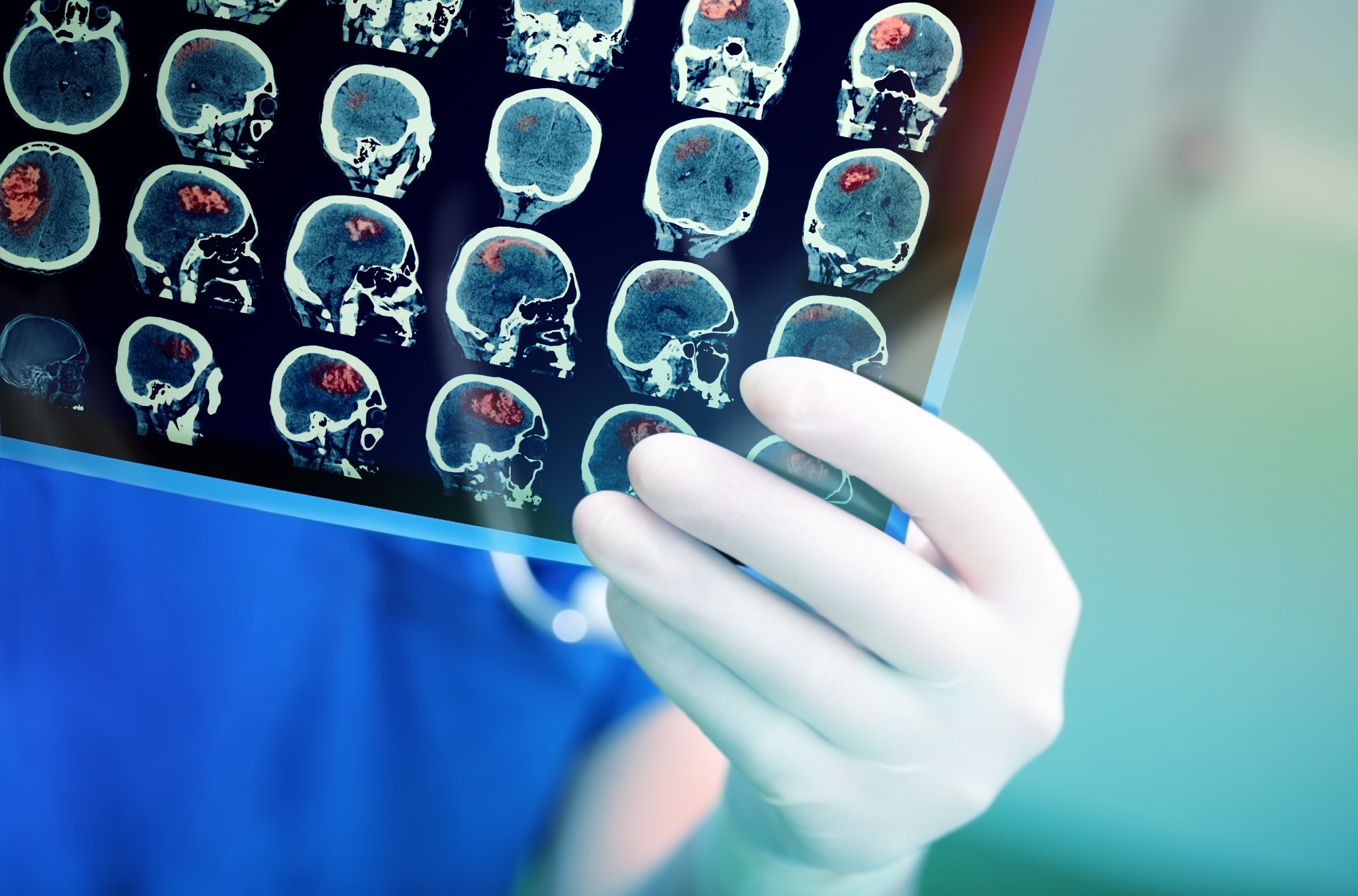
In a recent study published in the journal Brain, researchers reported that brain injury is common in coronavirus disease 2019 (COVID-19) and influenza.
COVID-19 has been linked to neurologic complications such as stroke, autoimmune encephalitis, and Guillain-Barré syndrome. While physical brain injury is evident in COVID-19-related neurologic syndromes such as encephalitis and stroke, various reports suggest COVID-19-associated brain injury could occur even in the absence of a concomitant neurologic diagnosis. In addition, the exaggerated inflammatory response during COVID-19 might drive progression to severe disease.
 Study: Brain injury in COVID-19 is associated with dysregulated innate and adaptive immune responses. Image Credit: sfam_photo / Shutterstock
About the study
In the present study, researchers investigated host markers of the dysregulated immune response. Polymerase chain reaction (PCR)-positive COVID-19 patients admitted to Cambridge University Hospital between March 2020 and March 2021 were included. Additionally, the patient cohort was supplemented with a convenience sample of COVID-19 patients from Sahlgrenska University Hospital, Sweden.
Healthy subjects were recruited before the COVID-19 pandemic as controls. Stored clinical and plasma data of influenza patients from a separate trial were used as an additional control cohort. A small positive control group was included as a reference for the magnitude of elevations of brain injury biomarkers. This group comprised patients with acute severe traumatic brain injury.
Serum samples were collected from admission and convalescence at up to three-time points – acute (< 14 days), sub-acute (15 to 70 days), and convalescent stages (> 80 days; outpatient). Influenza/COVID-19 patients were divided into three severity groups (mild, moderate, and severe) based on treatment during the acute phase. Mild patients did not require oxygen supplementation, moderate patients required supplemental oxygen, and severe patients needed mechanical ventilation.
Glial fibrillary acidic protein (GFAP), neurofilament light (NfL), and total tau concentrations were measured in the sera from COVID-19 patients or plasma from influenza patients. Autoantibodies were screened using a customized central nervous system protein microarray. Further, serum concentrations of tumor necrosis factor (TNF)-α, interleukin (IL)-1β, IL-10, interferon (IFN)-γ, and IL-6, were quantified using multiplexed particle-based flow cytometry.
Findings
The researchers obtained 250 samples from 175 COVID-19 patients and control samples from 59 healthy subjects and 45 influenza patients. Seventy COVID-19 patients had mild disease, 72 had moderate disease, and 33 patients had severe disease. The concentrations of GFAP, total tau, and NfL were above the functional lower limit of quantification for most healthy control and COVID-19 patient samples.
Notably, serum concentrations of GFAP and NfL increased with COVID-19 severity at acute and sub-acute time points, consistent with the levels observed in subjects with severe traumatic brain injury. No differences were observed in total tau levels between patients and controls. Longitudinal samples were available for 67 patients, which allowed for studying temporal dynamics.
The authors noted that NfL and GFAP concentrations declined with time, albeit some patients showed increased NfL concentration between acute and sub-acute periods. Serum GFAP concentrations were not different between patients and controls at convalescent timepoint. Nevertheless, serum NfL concentrations at convalescent timepoint were higher in patients with moderate or severe disease than in controls.
The increase in total tau concentration did not vary with disease severity. Convalescent levels of serum GFAP and NfL correlated with paired samples collected between 15 and 42 days, whereas total tau concentrations did not correlate. Moreover, GFAP and NfL concentrations in the plasma collected from influenza patients with severe disease were elevated to levels comparable to COVID-19 patients.
COVID-19 patients exhibited obvious IgG reactivity to spike and nucleocapsid proteins of severe acute respiratory syndrome coronavirus 2 (SARS-CoV-2) and, notably, to the lung surfactant protein A (SFTPA1). Reactivity to SFTPA1 was higher in sub-acute samples of moderate and severe patients than in mild patients or healthy controls. This autoantibody has not been described in the context of COVID-19.
Further, autoantibody profiles of cohorts were compared by determining the number and targets of positive autoantibody hits to specific antigens. COVID-19 patients had higher IgG and IgM autoantibody hits than healthy controls. Anti-myelin-associated glycoprotein (anti-MAG) was the most common IgG autoantibody, followed by anti-SFTPA1 autoantibody detected in 9.6% and 8.8% of COVID-19 samples, respectively. Both autoantibodies were not detected in healthy controls.
Increased serum cytokine levels were observed in sub-acute samples, and many convalescent samples had elevated cytokine concentrations above the normal range. Moderate and severe COVID-19 patients had elevated levels of proinflammatory cytokines. Next, the team investigated the associations between brain injury biomarkers and inflammatory profiles (cytokine and autoantibody responses).
There was a positive correlation between serum NfL and GFAP levels and the number of IgG hits. Nonetheless, total tau concentrations were not associated with IgG hits or cytokine profiles. The number of IgM hits was correlated with serum NfL levels but not with total tau or GFAP concentrations. Of note, the researchers found a correlation between the number of IgM hits and all brain injury biomarkers, particularly total tau levels, at convalescence.
Conclusions
The present study demonstrated elevated concentrations of brain injury biomarkers in COVID-19 patients, which increased with disease severity during acute infection. These elevations correlated with the presence of autoantibodies and proinflammatory cytokines. In addition, there was evidence of a dysregulated immune response even after four months. Notably, autoantibodies against brain antigens did not predict brain injury any stronger than those against non-brain antigens.
This meant that brain injury occurred due to general dysregulated immunity and not because of directly pathogenic autoantibodies. Furthermore, data from influenza patients indicated that brain injury during acute SARS-CoV-2 infection was not unique to COVID-19. Overall, the findings revealed the association of brain injury biomarkers with dysregulated immunity in COVID-19.
- Needham EJ, Ren AL, Digby RJ, et al. Brain injury in COVID-19 is associated with dysregulated innate and adaptive immune responses. Brain, 2022. DOI: 10.1093/brain/awac321, https://academic.oup.com/brain/advance-article/doi/10.1093/brain/awac321/6692467
Posted in: Medical Research News | Medical Condition News | Disease/Infection News
Tags: Autoantibodies, Brain, Central Nervous System, Coronavirus, Coronavirus Disease COVID-19, Cytokine, Cytokines, Cytometry, Encephalitis, Flow Cytometry, Glycoprotein, Guillain-Barré Syndrome, Hospital, Immune Response, immunity, Influenza, Interferon, Interleukin, Microarray, Myelin, Necrosis, Nervous System, Neurology, Oxygen, Pandemic, Polymerase, Polymerase Chain Reaction, Protein, Respiratory, SARS, SARS-CoV-2, Severe Acute Respiratory, Severe Acute Respiratory Syndrome, Stroke, Syndrome, Traumatic Brain Injury, Tumor, Tumor Necrosis Factor

Written by
Tarun Sai Lomte
Tarun is a writer based in Hyderabad, India. He has a Master’s degree in Biotechnology from the University of Hyderabad and is enthusiastic about scientific research. He enjoys reading research papers and literature reviews and is passionate about writing.
Source: Read Full Article
